Stress Fractures
Stress fractures can occur for many different reasons; for example over training or metabolic reasons.
There are two groups of bone cells that are responsible for bone turnover. One group breaks bone down osteoclasts and the other builds it up osteoblasts. Both the bone reduction and bone production needs to be in harmony with each other when that balance is disrupted, the rate of break down overrides the rate of build-up and the bone can become weakened. This is the mechanism of osteoporosis and osteopenia as well. Typically, this occurs in the second and third toes.
Symptoms of stress fractures include tenderness over the middle or just behind the toes on the metatarsal (long foot bone), which can produce moderate swelling on the top of the foot.
Any pressure or weight bearing can be very painful, and often people will have continued pain with rest. Initial X-rays are negative because the stress fracture is so small.
A good ortho will take repeat films at three to four weeks to see if the body has started to develop a callous along that stress fracture. If that isn’t visible and pain persists, a bone scan or MRI may be warranted.
Stress fractures are best managed with ice, decreased loading or non-weight-bearing, and casting in severe cases.
The bones of the foot need to absorb external forces from our day to day life style choices like walking, running, jumping or sporting activities you may choose to partake in.
When considering the amount of weight or forces applied to your bones and muscle we need consider that ground reaction forces increase not only with mass but with acceleration and deceleration.
Up to 12 times the weight of the body may be generated with each step; and the bones, joints, muscles, and ligaments need to cushion the body against that force. With that in mind we can now start to understand why even a small fracture in the foot of leg can be extremely painful.

Symptoms of a stress fracture may include pain and swelling, particularly with weight bearing on the injured bone.
Stress fractures commonly occur in the following body locations:
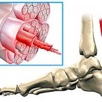
- metatarsal bones of the foot
- navicular bone in the foot
- calcaneus (heel bone)
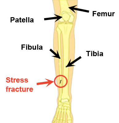
- tibia fibula (shin bones)
- femur (thigh bone)
Bone is normally in a balance or homeostasis (homeo= same + stasis=standing still), meaning that the natural turnover of bone cells is in balance between osteoclast activity (bone breakdown) and osteoblast activity (bone creation).
When bone is under excessive load or experiences trauma, microscopic damage can be caused.
Osteoclast cells are stimulated to absorb bone the damaged bone from the injured site. Providing sufficient time has passed before the next injury occurs, osteoblast cells produce more bone cells to heal the damaged area.
If there is not enough time for the osteoblasts to produce more bone cells in the injured area; the micro fractures can join together to form a large enough area to cause a stress fracture.
What to Learn More?
Interested to learn more about stress fractures or want to address your stress fracture with exercise physiotherapy? Get in touch with the physiotherapy Brisbane team from Pivotal Motion Physiotherapy on 07 3352 5116 today for physio with an integrated approach.
Updated 12/03/2021